Doctor Schedule
Clinics & Centers
See all
 Pricing
Pricing
Pricing
Programs & Packages
Promotions
Inpatient rooms
Programs & Packages
Promotions
Inpatient rooms
Ask for more

 Doctor Schedule
Pricing
Pricing
Doctor Schedule
Pricing
Pricing

.
The Radiology Department at Synphaet Hospital Srinakarin offers comprehensive radiology services with modern standard methods, enhancing efficiency.
Medical Technology
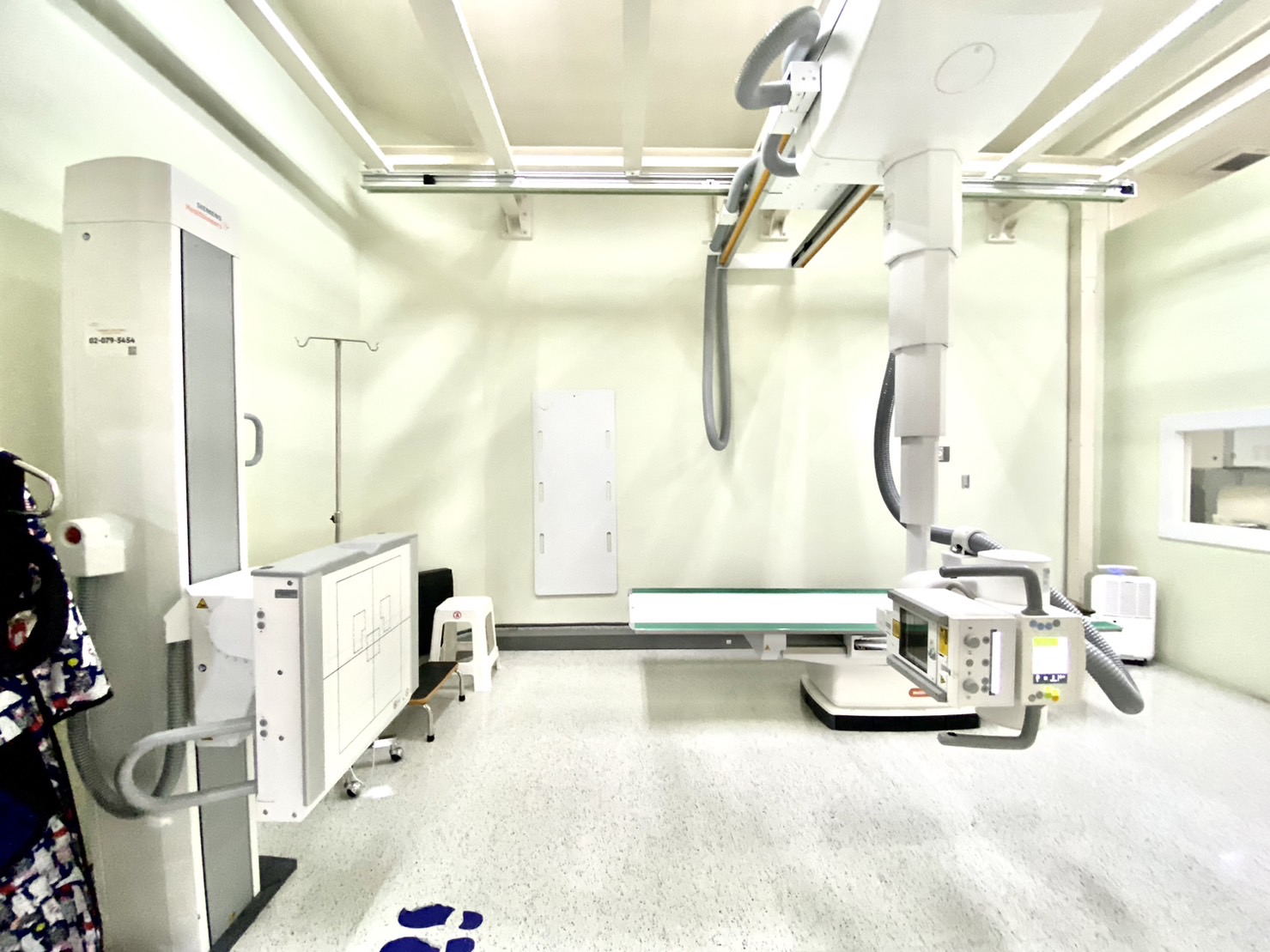
1.Digital Radiography (DR) is an imaging technology that uses computer systems to create images. The image is displayed in the computer’s memory and can be manipulated to focus on specific areas, enlarge, or adjust the opacity to study various densities like tissues or bones. This system reduces the patient’s radiation exposure compared to traditional film radiography and allows for data to be retrospectively viewed. It can perform general X-rays, such as chest, skull, arm, leg, and spine, which help in preliminary diagnosis of abnormalities, speeding up the X-ray process with images appearing within 3 seconds, providing clarity, and reducing repeated exposures.
2.Portable X-Ray machines are used for patients who are immobile or unable to assist themselves, or who cannot come to the radiology department, such as in the ward, emergency room (ER), intensive care unit (ICU), and operating room (OR). They can be conveniently moved to various required areas.
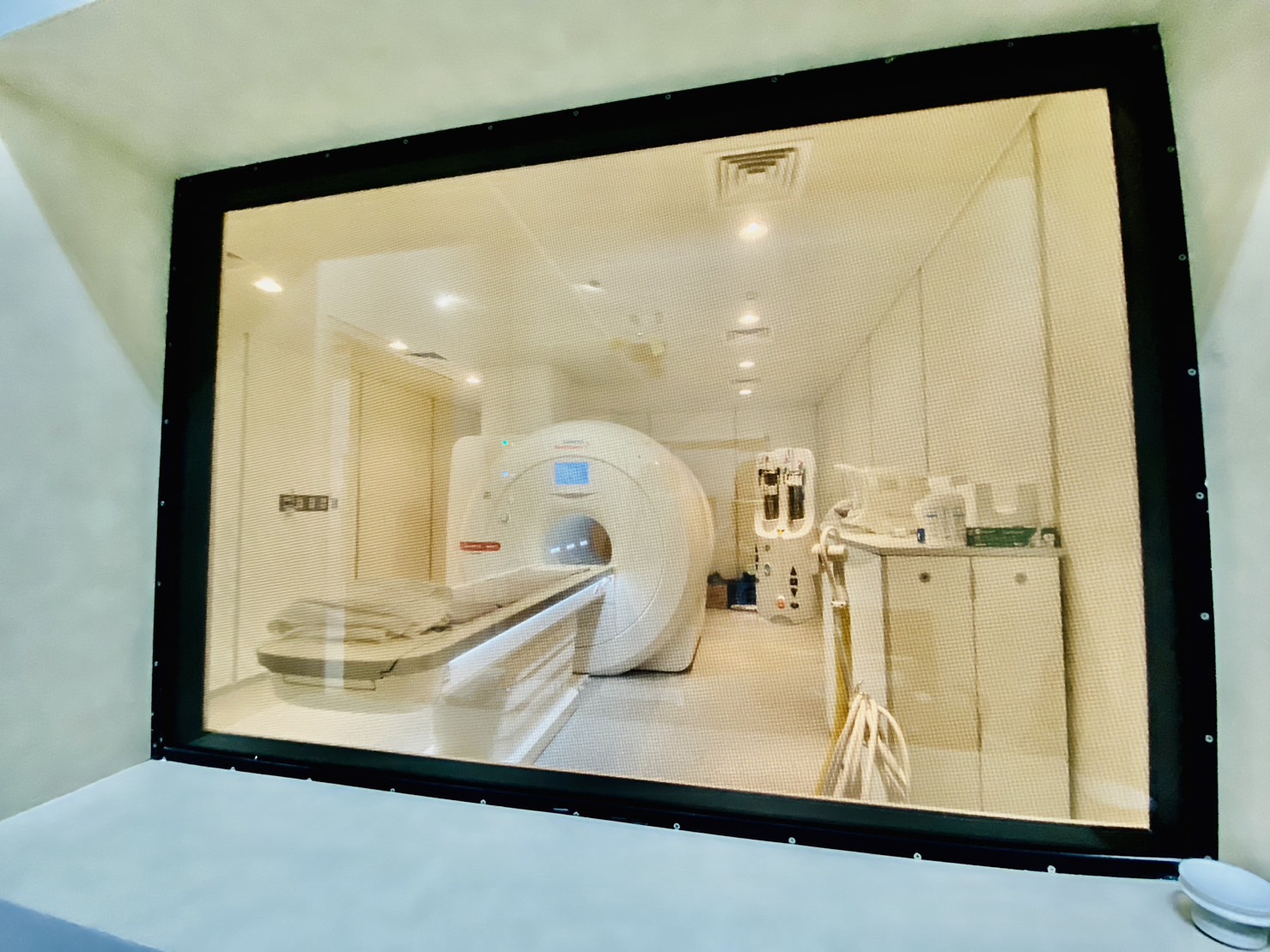
3.MRI (Magnetic Resonance Imaging) is a non-invasive medical device that uses magnetic and radio frequency waves to react with protons in different tissues, creating an image signal converted to internal organ images by a computer system. MRI does not use X-rays and hence is free from radiation exposure.
-MRI can detect brain abnormalities and nervous system issues such as strokes, tumors, causes of seizures, and brain and meningeal infections. It can identify abnormalities in abdominal organs, uterus, and ovaries in females, and the prostate in males.
-MRI is effective in diagnosing bone and joint abnormalities, spinal nerve issues like slipped discs, spinal tumors, infections, and spinal injuries. It helps detect muscle tissue abnormalities, including muscle tears, tendon injuries, and more.
-MRI is capable of visualizing blood vessels and blood flow abnormalities.
-MRI provides clear differentiation between various tissues, leading to more accurate and precise disease diagnosis. It can conduct examinations in all planes, not just cross-sectional like CT scans, and is especially effective for non-bony parts like soft tissues, particularly the brain, spinal nerves, and body nerves (CT scans better visualize bones). Additionally, it works well for muscles, tendons, and muscles.
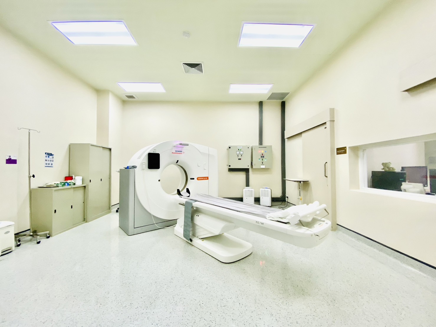
4.CT SCAN (Computerized Tomography Scan)A diagnostic procedure using a computerized axial tomography device, where the doctor will direct X-ray radiation to the specific area of the body that needs examination. A computer then generates images depicting the characteristics and internal organs of the body to aid in diagnosing any abnormalities. This method produces more detailed images than standard X-ray examinations and can be used to inspect nearly every internal organ.- Diagnose illnesses, such as detecting injuries to internal organs, internal bleeding conditions, blood circulation, thrombosis, bone fractures, ischemic brain conditions, tumors, and cancers.- Monitor the treatment of illnesses, both during and after treatment, such as checking the size of a tumor or the results after cancer treatment.
– Assist as a guideline for treatment, for example, determining the size and shape of a tumor before radiation therapy, using CT Scan to image while the doctor drains abscess fluid with a needle, or retrieves a tissue sample for examination.
– The examination takes approximately 45 minutes to 1 hour.
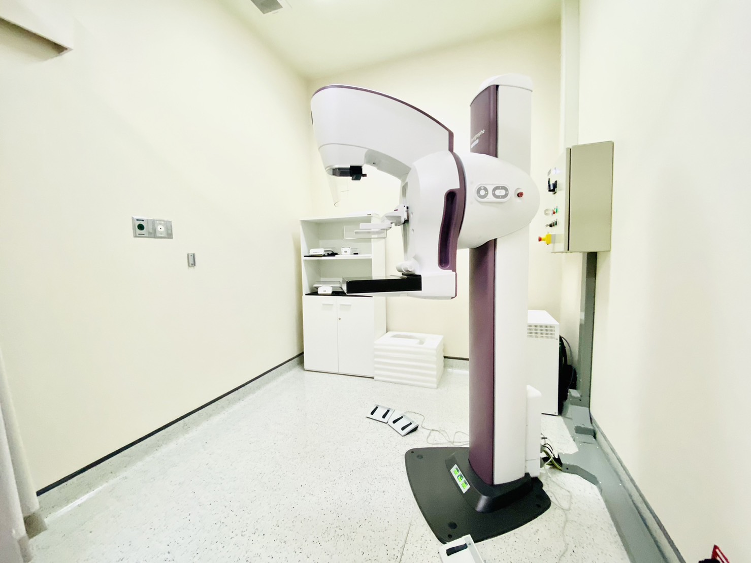
Digital Mammogram is a leading technology for breast cancer detection in the country, enabling Digital Mammograms to detect the disease from an early stage before symptoms appear, which may not be detectable during general health checks. With precise systems, it can efficiently examine tissue details. It is evaluated together with Ultrasound for better diagnostic outcomes for breast cancer. The Digital Mammogram examination is safe and has no harmful effects on the body, taking only about 10-15 minutes. Since breast cancer is a disease that cannot be detected by general health screening and does not show symptoms in the early stages, it should be diagnosed through a Digital Mammogram, a specific device for accuracy only. Early detection of breast cancer can make it easier to treat than at later stages due to the high quality, clarity, and precision of the images.
Ultrasound is a medical examination that uses high-frequency sound waves that penetrate into the internal organs to capture images of organs or various parts inside the body. Besides being used for prenatal baby checks, it can also be used to diagnose diseases or assist doctors in seeing the body while administering injections. It is a safe examination because it does not use X-ray radiation, magnetic waves, or radio waves, and does not involve injecting dyes. Therefore, it can be performed more frequently than X-ray and MRI examinations. It can examine every tissue or organ similar to X-ray and MRI examinations. Although the image resolution is not quite as high, it can still evaluate and diagnose effectively. It captures clear images of soft tissues better than X-ray examinations. The cost of examination with Ultrasound diagnostic is significantly less than X-ray and MRI examinations.
What can be diagnosed?
Tendons in various areas such as the shoulder tendon, elbow tendon, anterior knee tendon, etc.
Muscle fibers that are in pain to assess the severity and treatment direction
Various joints, such as knee inflammation, to assess whether the knee has swelling from fluid or how much degeneration and calcification have occurred, and used in the injection of various drugs to be accurate, precise, and safe in drug injection, such as Prolotherapy injection, Steroid injection, PRP injection.
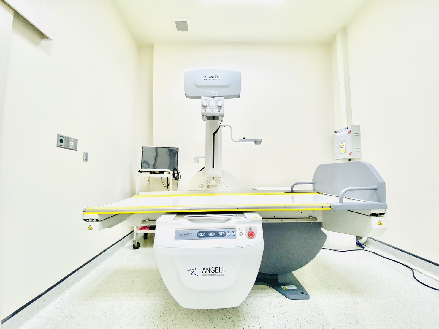
X-ray Fluoroscopy is an X-ray device used in diagnosis by radiologists to find abnormalities in internal organs in real-time, including examinations of the stomach and upper small intestine, and can record images of the movement of various organs, such as breathing, swallowing of the esophagus. The system consists of an X-ray table and an X-ray axis which is a C-shaped arm with an X-ray source under the table and an image receiver above the table, transmitting images immediately to the computer screen.
Bone mineral density (BMD) equipment to assess the risk of developing osteoporosis and the risk of bone fractures resulting from osteoporosis over a 10-year period.