




Detect Heart Abnormalities with a Cardiac MRI
Today, many people die from heart failure or sudden cardiac arrest. These conditions can occur from middle age through older adulthood. Risk factors vary, from internal factors such as family genetics to external lifestyle behaviors that can lead to heart failure.
A key prevention strategy is to get regular health checkups to reduce risks that could lead to future fatal events. Today, Synphaet Ramintra introduces how to detect abnormalities using a Cardiac MRI to help prevent, treat, and rehabilitateheart disease symptoms
What is a Cardiac MRI and how does it relate to heart testing?
A Cardiac MRI is a heart examination that uses magnetic resonance imaging. It produces highly detailed images of the heart’s structures, enabling physicians to evaluate cardiac function effectively—blood flow within the heart and problems in the chambers, myocardium, and valves—without causing pain to the person being examined.

What conditions can Cardiac MRI identify?
Cardiac MRI can identify diseases related to the cardiovascular system, such as:
- Heart failure
- Ischemic heart disease
- Cardiomyopathies
- Valvular heart disease
- Congenital heart disease
- Aortic diseases such as aneurysm, rupture, and dissection
When should you consider a Cardiac MRI?
Indications for Cardiac MRI are assessed by a specialist and may include purposes such as:
- Diagnosing causes of disease, e.g., acute myocardial ischemia or myocarditis
- Detailed assessment of cardiac structure and function, such as blood flow in great vessels entering the heart
- Evaluating treatment results before and after surgery
Cardiac MRI procedure
A Cardiac MRI involves three main stages:
Before a Cardiac MRI
Inform your doctor beforehand if you are pregnant or may be pregnant; have a pacemaker, an intrauterine device, or other implanted medical devices; or if you have severe claustrophobia. Your doctor may prepare a sedative or provide guidance to ease anxiety.
During a Cardiac MRI
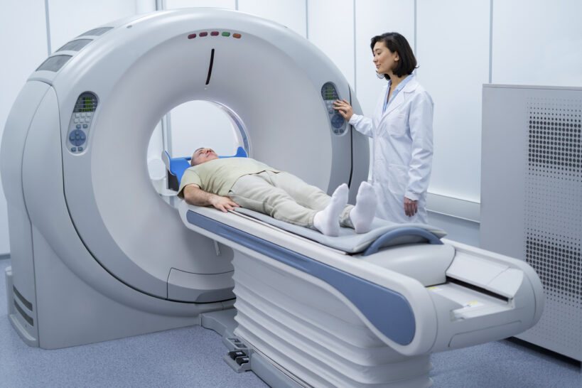
You will lie on a table that slides into the MRI scanner. Radiofrequency waves target the chest to create detailed images of heart tissues and coronary vessels. A contrast agent may be injected intravenously to enhance clarity. The scan typically takes about 30–90 minutes.
After a Cardiac MRI
Your doctor will schedule a review of the results. Most people can resume normal activities without recovery time. If contrast was used, you may briefly experience a bitter taste, dizziness, or nausea; resting until you feel better is recommended before going home.

Benefits of Cardiac MRI
Key advantages of Cardiac MRI for evaluation and diagnosis include:
- Detailed assessment of cardiac structure, function, and blood flow
- Detection of structural abnormalities—valves, myocardium, and pericardium
- Diagnosis of cardiac and vascular diseases, such as coronary artery stenosis or occlusion
- Evaluation of vascular-related damage, such as restricted blood flow to the heart
- Informing and planning patient treatment
- Monitoring disease progress, including post-operative changes
Who may not be suitable for Cardiac MRI?
Although Cardiac MRI is accurate, effective, and safe, it may be unsuitable for some groups:
- Pregnant individuals or those who may be pregnant
- People with implanted medical devices, such as pacemakers or defibrillators
- People with metallic implants, e.g., metal clips, mechanical heart valves, or cochlear implants
- People with severe claustrophobia
- People unable to lie flat
Summary
Cardiac MRI provides precise, detailed evaluation of cardiac function and blood flow without causing pain. It supports prevention, treatment, and rehabilitation of heart disease effectively. We hope this article encourages ongoing health checkups for long-term wellness and disease prevention.